Tag: Yale Fisher

  • Glaucoma Awareness: Early Detection Saves Sight

    Glaucoma Awareness: Early Detection Saves Sight

    January is Glaucoma Awareness Month, strategically placed at the start of each new year to remind us to schedule our annual comprehensive eye exam because early detection is still the best way to prevent the irreversible vision loss that can be caused quietly in the early stages and without noticeable symptoms.  According to Prevent Blindness, more than 2.8 million Americans age 40 plus have glaucoma and nearly half do not know they have the disease.

    When it comes to glaucoma awareness, it’s always a good time to improve your understanding and evaluate your risk.

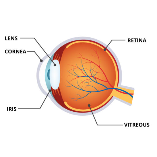
    Glaucoma is an increased pressure in the eyeball. It can be thought of as a “plumbing problem” with either too much fluid entering a closed system or not enough fluid going out. The result is an elevation in the intraocular pressure. Over time the pressure will irreversibly damage the optic nerve, which sends visual information from the retina to the brain.

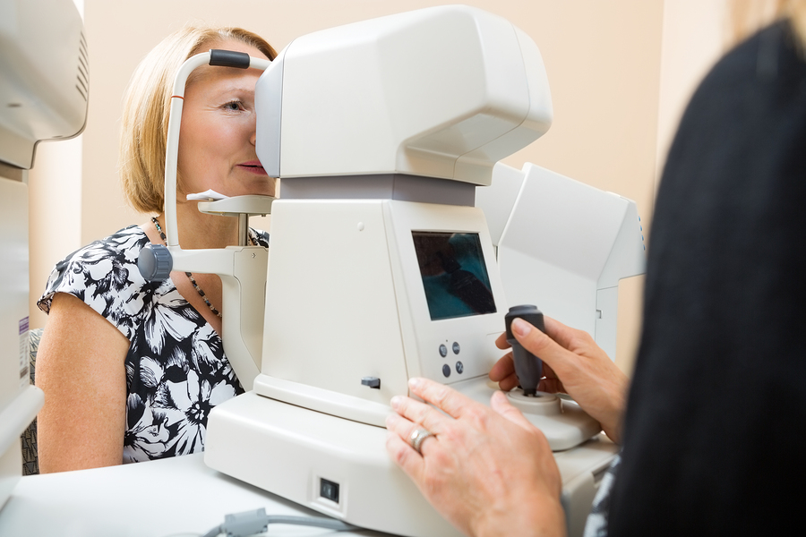
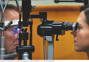
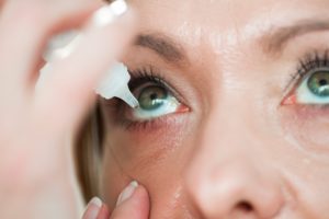
    There are no overt symptoms, so the vision loss which begins peripherally can quietly progress unnoticed. Early detection is key to saving sight. Regular comprehensive eye exams are the best way to detect glaucoma in its early stages. Although there is no cure, there is effective treatment. Eye drops are commonly prescribed to control the pressure. 

    You are at a higher risk for developing glaucoma if you have a family history (especially a sibling), are of African, Asian or Hispanic descent; are over age 60, have diabetes or are severely nearsighted. If you are affected by any other eye disease it is also important to have your pressure tested regularly.


    Woman giving man eye pressure test
    Woman giving man eye pressure test. Credit: Mayo Clinic

    It should also be noted that pressure problems often occur after retinal procedures and may last for short or long periods of time.  Dependent on individual patient anatomy or pathology, these pressure changes require careful and sometimes immediate attention if permanent damage to the optic nerve is to be curtailed or prevented.  Pressure elevations may even occur long after retinal procedures, a result of “altered” slow changes in tissues over time. While everyone should be aware of the need to check their intraocular pressure on a routine basis, those having undergone a retinal procedure should be especially vigilant in the immediate postoperative period and periodically thereafter.  Often slow elevations occur without noticeable symptoms.

    Should you find yourself in any of the risk categories mentioned, please consult your ophthalmologist and be meticulous in keeping up with your exams to avoid visual impairment, legal blindness, and potentially even total blindness.

    Eyes with dropper
    Eyes with a dropper. Credit: RD.com


    This article has been updated from original version published January 11, 2018.

  • Doctor, Am I Going Blind?

    Doctor, Am I Going Blind?

    To the question, “Am I going blind?,” the answer is most often, “No.”

    For the great majority of patients diagnosed with disease-causing progressive vision loss, use of the word “blindness” actually does not apply.  Fortunately, most patients treated for degenerative diseases retain some degree of visual function.

    While the amount varies (dependent on disease type, severity, persistence, and progression), it is far more accurate to describe most cases as visual impairments and not as blindness.

    Obviously, total loss of light perception is the ultimate fear.  Ophthalmologists must be extremely careful in their discussions with patients to address this fear.  While some diseases are severe, most can be limited.

    Macular Degeneration

    A frightening diagnosis always, and is the most common ocular disease in older age.  Some forms are genetically inherited and present in adolescents, teens, and much younger adults.  Although often described as the “leading cause of blindness in people over 65,” it is highly unlikely and extremely rare that it results in loss of light perception or blindness.  Macular degeneration generally affects the central vision, leaving peripheral intact.

    Progression of the disease is slow in most cases and can be well compensated.  Patients are able to continue their full and productive lives, making some adjustments and utilizing magnification and speech in widely available accessible technologies.  A strong mental approach coupled with a willingness to learn, are proven factors in maximizing remaining vision and enjoying life.

    Diabetic Retinopathy

    Diabetes-related eye disease is another common malady often associated with the reduction in normal sight.  While historically unrelenting, attention to medical and ocular problems early in the course of diabetes have completely altered the potential for devastating visual loss.  With current and updated treatments, visual loss can be prevented or at least limited.  Often, patient cooperation and participation in treatment is the key to success for diabetics.

    Glaucoma

    Primarily a disease of superficial retinal loss associated with elevated pressure within the globe of the eye.  The earliest patterns of visual loss from glaucoma are peripheral, not central, so a reduction of visual field can go relatively undetected by the patient.  As the disease progresses, more of the peripheral field is lost, followed finally by the loss of the central areas.

    Fortunately, with treatment and early recognition by a qualified professional, visual loss can be prevented or limited.

    Legal Blindness

    Many have heard the term “legal blindness” and automatically interpret it to mean “blindness.” Legal blindness is a government definition or statute utilized to determine qualification for disability benefits.  The definition requires 20/200 vision or worse in the best eye with correction in place or visual field limitation to 20 degrees in diameter.  The condition causing the vision loss must be present or expected to be present, for one year or more.

    Legal blindness does not mean “no vision” and many people with this degree of vision loss live very full visual lives, albeit with a significant visual impairment.  On the other hand, “blindness” is usually understood to mean “no visual perception”.

    Clarification of the language associated with vision loss is critical, especially for those on the receiving end of these difficult words.  Speak with your ophthalmologist.  Make sure you understand your visual status and to what degree it is likely to change over time.  Most of the time vision can be preserved and loss can be limited.

  • Should Floaters and Flashes Concern Patients?

    Should Floaters and Flashes Concern Patients?

    To understand why floaters and flashes happen, you first need to review a little bit of eye anatomy.

    The eye is essentially a ball which I like to divide into three areas of interest.  The front (or cornea) is the window that allows light into the eye.  The middle is composed of the iris, pupil and focusing lens.  Behind the iris-lens area is a large cavity filled with a clear gel-like substance called vitreous.  And behind the gel is the back wall that is lined with a thin “wallpaper” called the retina.

    Most do not realize that the retina is actually an extension of the brain.  So retina is really brain tissue!  It is responsible for transforming light that enters the eye into electrical signals that are processed and interpreted by the rest of the brain into shape and color.  You really do not see with your eye.  The eye is really a transducer of energy, light into electrical signals.  The rest of your brain forms the image.

    Anatomy of an Eye
    Anatomy of an Eye

    Why is the doctor so interested in FLASHES and FLOATERS?

    The usual normal process of vitreous gel aging and separation from the retina occurs over a lifetime and does not usually cause problems.  The gel liquefies and collapses separating from the retina.

    During this process, the patient may begin to notice small translucent or darker particles occasionally “floating” in their field of view.  These floaters represent the projected shadows of fine vitreous particles floating in front of the retina.  In some situations, however, the separation of the gel occurs too suddenly or the pull of the gel is too forceful for the delicate retina resulting in retinal tears.

    Usually, these more violent vitreous separations are associated with more symptoms- more noticeable flashes and floaters.   (The retina does not have pain fibers only light fibers so when pulled or torn, light flashes are seen).  Doctors take all flashes seriously.  With careful examination, it is possible to determine if tears occur and treat damaged areas before any further changes follow.

    Often a tear may be associated with many floaters, not just a few minor shadows, but large clumps.  These larger and more numerous floaters often represent the clotted blood that exudes from vessels at the edge of the torn retina.  These clots absorb over time, the floaters appear to decrease.  The tear or tears do not repair themselves and some permit liquid to invade beneath the retina producing first a blister of retinal tissue around the tear(s).

    With time and eye movement, more and more fluid infiltrates under the retina creating a larger separation of the retinal “wallpaper”.  When larger separations are present they are called retinal detachments.  A detached retina is seen by the patient as an increasing shadow or veil-like black area within the normal field of view.  Detachments require more extensive treatment (often surgery) to reattach the separated areas.

    So floaters may be simply a normal aging process (common) or a symptom of something more ominous.  Most individuals go through the process of vitreous gel separation without tear formation, however, it is always prudent to have a careful exam until the gel separation is complete and the symptoms subside.

    Strong flashes and many floaters are more alarming symptoms and are more often associated with damage that may require immediate attention.  Early detection is the best way to improve the chances for success in treating retinal tears and avoiding detachments with potential serious vision loss.

    An examination is always better sooner than later.