Category: Health & Well-Being

Pay attention to your health & well-being with expert insights on eye health and vision care.

  • Treatment For Diabetic Retinopathy

    Treatment For Diabetic Retinopathy

    If you have diabetes and develop any changes in your vision, contact your ophthalmologist immediately.  While we currently do not have therapies that can completely reverse diabetic retinopathy (DR), we have effective treatments that can slow down or stop its progression and prevent your vision from further deteriorating.  In some cases, therapies are effective at recovering some lost vision.

    Therapies are employed to treat swelling of the central retina called diabetic macular edema (DME) and for treatment of severe DR that compromises blood flow to the retina.


    Diabetic Macular Edema

    If you are experiencing DME, your doctor might suggest injections into your eye with a medication known as anti-vascular endothelial growth factor (anti-VEGF) agents.  These medications help to decrease the macular edema and improve vision.  While the idea of a needle being inserted in your eye scares almost everyone at first, these therapies are highly effective and are in fact, a common procedure that retina specialists perform daily in their office.  There are multiple anti-VEGF medications to choose from for diabetic macular edema.  You should discuss with your retina specialist to determine which one is most appropriate for you.

    In some cases, your doctor might also recommend an injection into your eye of an anti-inflammatory medication known as a steroid.  These injections may be suggested in patients that have had a prior cataract surgery and are experiencing worsening of their DME or have chronic DME despite treatment with anti-VEGF injections. 

    If the DME is just outside, but threatening to disrupt the central vision, your doctor may recommend a light focal laser to seal off the leaking blood vessels that are contributing to the retinal swelling.


    Diabetic Retinopathy illustration diagram.

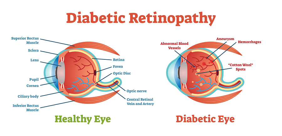
    Severe Diabetic Retinopathy

    If your DR results in a severe compromise of blood flow to the eye, your doctor may recommend a laser called panretinal photocogulation.  This laser, while sometimes painful or uncomfortable, is remarkably effective in preventing total blindness from bleeding in the eye and retinal detachment. 

    If there already is blood in the eye, with or without scar tissue on the retinal surface, your doctor will likely recommend a surgery known as a vitrectomy to remove a gel-like substance inside your eye called the vitreous body.  This procedure can also remove the bleeding and some of the scar tissue that has developed.  These treatments are complex and visual recovery expectations need to be discussed with your treating retinal surgeon.

    Recently, there has been growing evidence to support treatment of DR with anti-VEGF injections.  While these treatments are effective, the frequency of treatments could pose compliance challenges for patients.

    In conclusion, several management and treatment options are available for patients with diabetic eye diseases.  Please consult with your doctor on the best management plan for you.


    Written by:

    Luis Martinez-Velazquez, MD, PhD will be starting his ophthalmology residency at Massachusetts Eye and Ear Infirmary.
    Yasha Modi, MD is an Assistant Professor of Ophthalmology at New York University specializing in vitreoretinal surgery, retinal disease, and uveitis.


  • What Is Diabetic Retinopathy?

    What Is Diabetic Retinopathy?

    If you have been diagnosed with Diabetic retinopathy (DR), you’re not alone.  DR is a common complication of both type 1 and type 2 diabetes and a major cause of vision loss in adults of all ages in the United States and throughout the world.  One-third of the people with diabetes have DR, that translates to an astonishing 95 million patients with diabetic eye disease worldwide. 

    How long does it take for diabetes to damage eyes?

    Studies done in the United States have shown that patients with diabetes typically develop some form of DR within 10 years of diagnosis.  While some forms of DR are quite mild, one-third of patients will develop vision-threatening retinopathy.

    Diabetic eye disease is caused by damage to the blood vessels and neurons in your retina, the light-sensitive tissue of your eye.  It is a progressive condition, meaning the longer you have diabetes and the higher your blood sugar, the more likely you are to develop this eye complication.

    What is the first sign of diabetic retinopathy?

    In the early stages, DR may not cause any symptoms or only mild changes in vision.  Nevertheless, even at these early stages, there is damage to nerve cells and blood vessels that your doctor would be able to see during a retinal examination. It is important to catch DR in its early stages before it progresses to the more advanced stages that threaten eyesight.

    At any stage of DR there could also be changes that lead to fluid accumulation, also known as edema, in the retina.  If fluid accumulates in the macula, or the central area of the retina which provides your sharpest vision, the vision may become severely compromised.

    How can you tell if diabetes is affecting your eyes?

    If you have diabetes, some of the symptoms that may indicate retinopathy include intermittently blurry vision, new floaters, and dark or blurry areas in your vision.  Because diabetes affects your whole body, these symptoms usually affect both eyes.

    Man with black eye glasses having difficult time trying to read book.

    Who should get screened for diabetic retinopathy?

    Patients with type 1 diabetes should have yearly eye examinations to screen for DR starting five years after their diabetes diagnosis.  Patients with type 2 diabetes should be screened starting the year of their diagnosis. 

    Women who develop gestational diabetes do not require an eye exam during pregnancy because studies currently indicate that they do not have an increased risk of developing DR.  However, women with diabetes who become pregnant should be examined early in the course of the pregnancy.  Timely detection can help control the disease before it can damage your vision.  All patients diagnosed with DR should be referred to an ophthalmologist, or more specifically a retinal specialist, to help manage the visual complications of diabetes.

    Can lowering blood sugar improve vision?

    Two of the most important steps you can take to preserving your vision are lowering your blood sugar and controlling high blood pressure.  In fact, good sugar control can often slow the progression of mild to moderate retinopathy.  Your doctors can also assist by monitoring and helping you control your glycosylated hemoglobin levels (A1C), serum lipids, and blood pressure.

    Is diabetic eye disease reversible?

    If you have diabetes and develop any changes in your vision, you should contact your ophthalmologist immediately.  While we currently do not have therapies that can completely reverse diabetic retinopathy, we have effective treatments that can slow down or stop its progression and prevent your vision from deteriorating further.  In some cases, therapies are effective at recovering some lost vision.


    Written by:

    Luis Martinez-Velazquez, MD, PhD will be starting his ophthalmology residency at Massachusetts Eye and Ear Infirmary.
    Yasha Modi, MD is an Assistant Professor of Ophthalmology at New York University specializing in vitreoretinal surgery, retinal disease, and uveitis.


  • Cataracts Removed, Vision Improved

    Cataracts Removed, Vision Improved

    For many years I asked my retina specialists (and there have been many) about the removal of cataracts.  For the most part they all said, “The risk isn’t worth the reward.”  Then finally, my current physician, Dr, Michael Tolentino, agreed it was time.  He said his perspective changed from that of caution and concern that the surgery could exacerbate the underlying macular degeneration.  He was beginning to see that patients who opted to have the cataract removal procedure were getting very good results in terms of clearer and brighter sight. 

    Dr. Tolentino recommended Dr. Melissa Iglasius, who he said was very competent and experienced in cataract surgery.  I interviewed the surgeon, and she gained my complete confidence.  Dr. Iglasius said definitively, “Standard surgery is sufficient, no need for laser or crystal lenses.”  There would be no additional out of pocket expense.

    I had both eyes done, within the last four weeks and the results are truly amazing.  Bear in mind that my visual acuity was 20/400 in the left eye and 20/200 in the right.  Before the surgery I could not see the letters on a ZoomText large print computer keyboard.  I can now see the letters clearly.  At this point I can also make out items in the fridge, that I could not see before.  The clarity in my peripheral vision is noticeably improved.  I still have macular disease, but the increase in light is a big step up.  The eye drops are to be continued in the right eye for 16 days, so there is potential to regain more clarity.

    Illustration showing normal eye on the left and eye with cataract on the right.

    When asked, at the support group meetings I run, about the removal of cataracts with AMD, my reply was always, “Discuss it with your retina specialist.”  The fact remains, a careful review of the possible risks and rewards of any medical treatment, should be addressed with your physician.  Today, based on my experience, I would also be inclined to encourage others to explore the options with an open mind.

    Cataract surgery is over in 15 minutes, recovery from anesthesia, depending on which one you chose, is relatively short.  It is well advised to adhere to the follow up instructions including a regimen of drops, no bending over, no rubbing the eye, and using anti-bacterial cleanser.  In my case, the reward was certainly worth the risk.  Thanks to Dr. Iglasius.


  • Grandpa, You Can See That?

    Grandpa, You Can See That?

    There is a famous proverb I learned in drama class many years ago and thought it apropos for this article.  “Do not judge a man until you’ve walked two moons in his moccasins.”  I offer up this advice, with a tinge of humor, for family, friends, colleagues and caregivers.  Having contracted macular degeneration more than 50 years ago, I feel well qualified to share my insights.

    Grandkids, or maybe even spouses, leave shoes, toys, and other obstacles in the middle of the living room floor.  They know you have a visual impairment, but it’s their house too, and this is normal behavior.  There are other usual and obvious hazards created by loved ones all the time, unintentionally.  Cabinet doors, closet doors, and drawers left open are undetectable by my eyes until it’s too late, and a collision is the end game.  The Bible tells us, not to place stumbling blocks in the path of the blind.  Although this may be a metaphor for life’s travails, for me it’s a truism.

    The pointing finger really gets my goat.  I walk into a retail store wearing the standard blindness indicators, dark glasses and white cane.  I ask a sales associate for help locating an item.  The answer comes back, “It’s over there,” and I see, peripherally, the finger is pointing in a general direction.  To avoid repeating this experience, I learned it’s best to immediately go to the register and state directly, “Can you help me find something? I’m blind.”  This pronouncement is necessary because in many stores the training is not inclusive of the accouterments of vision loss, or how to accommodate customers who are visually impaired.

    My lovely wife is always exhorting me to use the motorized cart when we shop, because I have difficulty walking.  I constantly tell her, “Honey, I’m blind in my left eye, leaving me with no depth perception, and I’m afraid I’ll hit somebody.”

    Neighbors, not accustomed to living with a person who has low vision, can be unaware of the pitfalls.  Serving me water in a clear glass is a risky move, placing the glass on a mirrored coffee table top, totally treacherous.

    Sometimes, even the youngest child can surprise you.  It happened on a trip to the zoo with our 5 year old great-granddaughter.  We were enjoying ice cream cones and getting a little messy.  My wife asked where the ladies room was and I pointed to the restrooms behind her.  My little one exclaimed, “Grandpa, you can see that?”  I told her that I knew where it was because I had used the facilities before, but was charmed at her cognition of my condition.

    And a child shall lead the way.


  • Healthy Aging And Eyes

    Healthy Aging And Eyes

    We develop many habits during the course of our lives, but not until we reach a certain age do we begin to understand their long-term effects.  The proof of our good and bad habits begin showing, if not in our physical appearance, they begin revealing themselves in our medical records.

    Maintaining overall good health is a key factor in preventing or slowing the progress of vision loss caused by age or genetics.  May is awareness month for Healthy Vision and for Older Americans, a perfect opportunity to reiterate, the many ways healthy aging is good for the eyes. 

    Take some of these steps today to preserve better sight for tomorrow.


    Fresh vegetables and fruits spread out on a gray table.

    Nutritious Diet

    This is your first line of defense against age-related disease and good habits can be developed at any time.  The earlier the better, so don’t delay.  Lower rates of heart disease, diabetes and age-related macular degeneration are directly attributed to diets abundant in colorful whole fruits, leafy green vegetables, fish, chicken, nuts, beans and whole grains.  Do your best to lower intake of refined sugar, saturated fat, sodium, and process foods in general.

    For more on OE, go to: The Anti-AMD Diet


    Happy couple wearing sunglasses outdoors.

    Sun

    UV rays are damaging.  Protect your eyes and your skin from the sun.  The earlier these habits begin, the better.

    For more on OE, go to: Keep The Sun Out Of Your Eyes


    Don’t Smoke

    The number one controllable risk factor for both AMD and heart disease.  Quitting today will significantly improve your health.


    Eye Exams

    A dilated eye exam can be the early detection that helps prevent or minimize vision loss from age-related macular disease, glaucoma, and diabetic retinopathy.  If you haven’t seen your eye doctor lately, it’s time to schedule that appointment, and keep them up on a regular basis, at least annually. 


    Happy mature man in a gym exercising.

    Exercise

    Regular exercise is a key ingredient for good overall health.  Several hours a week of moderate aerobic exercise and strength training are recommended for all adults.  Walking, hiking, swimming and dancing are also good options.


    Sleep

    Getting enough sleep is directly related to good mental, physical and emotional health.  It is quite possible you’ll see, feel, look and eat better following a restful night’s slumber.  And don’t let anyone tell you that older people need less sleep.  All adults require the same 7 to 9 hours per night.   


    Healthy Weight

    The combination of a nutrient rich diet and regular exercise, with good sleep habits, will help you maintain a healthy weight.  On the other hand, obesity has a long list of related illnesses, including diabetes and heart disease, which have a direct relationship to vision loss.


    Group of happy seniors laughing outside.

    Socialize

    The theme of this year’s Older American’s Month is “Connect, Create, Contribute,” and there is no question that the connection with family, friends and community is an essential element of healthy aging.


    A healthy lifestyle is all about balance. There are no magic formulas, create the mix that works best for you.  And remember, we are always a work in progress.


  • Don’t take the fall, prevent it

    Don’t take the fall, prevent it

    Falling is unpleasant at any age, for older adults it is downright dangerous.  Getting up from a fall becomes more difficult with each decade, and we are much slower to recover, both physically and emotionally.  There are many contributing factors that increase the risk, vision loss is certainly one of them.

    Hindsight is, as they say, 20/20.  Succumbing to injury is always regrettable, and in retrospect we often realize, it could have been avoided.  With the help of Alice Massa, an experienced Occupational Therapist and low vision expert, we’ve organized a list of guidelines to elevate your awareness about falls prevention and keep you on your feet.


    1. Vision

    Consult with your ophthalmologist to insure you are maximizing the vision you have.  Updating prescriptions and having cataracts removed can help make your surroundings a little more clear and easier to negotiate.


    1. Balance

    Remaining steady on your feed has a lot to do with good balance.  If you are feeling unsteady or faint, sit down immediately to avoid the fall. Regular exercise and strength training is highly recommended.  Each day practice standing on 1 leg to improve balance.  For more about the Single Leg Stance click here.  


    1. Floor Coverings

    Rugs and runners can be treacherous, be sure they are flush to the floor and have non-slip backing.  Get rid of rugs or carpet with fraying or curled edges.  Repair damaged or uneven flooring.


    1. Walkways

    There should be zero tolerance for clutter on floors.  No wires or cords that can catch a foot and take you down.  Something as innocent looking as a single sheet of paper can cause a deadly slip.  Shoes, particularly dark shoes on a dark floor, can go unnoticed, creating a serious hazard. 


    1. Footwear

    Wear well fitting shoes and boots with flat, non-skid soles.  If you do not feel secure in a shoe, get rid of it.  Walking around the house in socks, or barefoot, is not recommended, broken toes are also extremely painful and debilitating.


    1. Lighting

    Don’t skimp on lighting.  Make sure rooms are well lit and light switches are easily accessible.  Keep night lights on and a flashlight on hand.


    1. Kitchen

    Clean up spills immediately to prevent slips.  Keep frequently used items on lower shelves, to minimize the need for step stools.


    1. Bathroom

    Make sure your tub has a non-skid surface and grab bars if needed.  Keep a light on for travel to and from the bedroom during the night.


    1. Streets

    Cracked and uneven sidewalks create serious risk.  Pay attention and step carefully.  Using a cane can help you identify changes in the elevation of walkways and street crossings.  Read “Consider The Long White Cane” on OE.


    You know, they also say, an ounce of prevention is worth a pound of cure.  Take steps today to avoid the fall, and the regret.


  • A New Doctor, A New Course

    A New Doctor, A New Course

    As a member of Humana for many years, I have had to change my retina specialists several times.  I was very happy with the doctor I had for the past year and was surprised when I got a call, from my eye care center, informing me that I would now be cared for by a different retina specialist.  The woman on the phone said his name was Dr. Michael Tolentino, and that he had an excellent background and credentials.  Not taking her word for it, I Googled him.  I learned that indeed this was true, having been associated with the Schepens Eye Institute in Boston that I frequented in the 1980s.  So, I made my first appointment.

    He was very friendly and warm, and surprisingly, not Italian as his name would imply, but from the Philippines.  He told us that his father was a renowned ophthalmologist, associated with Schepens. He, himself had worked in the Schepens lab when he was 14 years old.  I learned that we both knew and respected many doctors that had trained there.

    Upon perusing my records and pictures, he said that he didn’t agree with my previous retina specialist’s decision to skip the eye injection at my last visit.  He showed me the pictures: “See that white part in both eyes, we have to get rid of that, and I’ll give you the shots until we can get your vision better.”  I was amazed to hear a doctor so aggressive.

    My next question to him was: “I have cataracts in both eyes, none of my previous doctors would allow me to have them removed. They said that the risk isn’t worth the reward, which would be minimal.’’  His response was: ‘’What risk? After a couple of shots, you’ll get both removed.”

    I remembered an article on cataracts from Lighthouse International a few years ago, stating that 20% of AMD patients had better visual acuity after cataract surgery.  I also wanted to be one of those 20%.

    Well, I put my faith and my eyes in the hands of Dr. Michael Tolentino and received my first 2 injections of Avastin, done very carefully and expertly.  I’ll keep my readers posted on the results.

    To learn more about cataract surgery and AMD from the American Academy of Ophthalmology…click here


  • Accessibility Matters In The Ophthalmologist’s Office

    Accessibility Matters In The Ophthalmologist’s Office

    A visit to any doctor’s office can include some degree of difficulty for people with uncorrectable vision loss.  The same challenges can even exist in the office of the ophthalmologist.  Good patient care is not just measured by the exam and treatment, it is also measured by the patient experience.

    Follow these guidelines to establish policies and procedures that help create an accessible office environment that is better for everyone.


    Make Sure Signage is Easy to Read

    Signs throughout the office should be in large print, using legible, highly contrasted easy to read font styles.  Never use print on a patterned background, or gold letters on a beige wall.  Positioning signs close to eye level makes them easier for everyone to see. 


    Utilize Color Contrast

    Use contrast in the office design.  Paint doors or frames a color that stands out from walls.  Use dark seating against a light carpet color.  Steps should always have contrasted edges and handrails. Avoid glass doors and walls entirely, they are hazardous to people with low vision and also those with dilated eyes. 


    Use Good Verbal Communication

    Always address patients directly by name, as they may not be able to see that you are speaking to them, or make eye contact.  When you greet a patient, identify yourself by name, don’t assume they will recognize you or know your voice.  Generally, it is not necessary to speak loudly to people with vision loss, unless you know their hearing is also impaired.

    Ask the patient if they need your assistance.  Don’t make assumptions about their abilities or their needs.

    Don’t point or say, “over there,” when giving directions.  Orient to the person’s direction by saying, “Walk to the end of this hall and turn left, the exit is the first door on your right.”


    Provide Accessible Information 

    Obviously, it is incorrect to assume all patients are able to complete paper forms.  If your office is still using clipboards to collect information, there should be alternatives offered.  Better options for everyone include online forms or having a staff member directly input all necessary information, provided by phone, pre-appointment.

    Make sure written communications are accessible.  Any printed or digital information given to patients should use text at least 18 pt., or larger and bolder when possible.  Always use a clean, easy reading, sans-serif font style like: Arial, Helvetica, or Verdana.  Be sure the content on copied documents is clearly legible.

    For many patients, particularly those who cannot easily read print, digital documents are the best alternative, as they can be adjusted to the reading preference of each individual.

    Remember, small changes can make a big difference in the way patients feel when they visit your office.


  • Hope For Age-Related Macular Degeneration

    Hope For Age-Related Macular Degeneration

    Age-related macular degeneration (AMD) is a common eye condition that impacts millions in the United States alone. In our previous article, we explored the different levels and types, as well as major risk factors.

    Thankfully, treatment is an effective option now.

    Even just fifteen or so years ago, having the advanced form of AMD almost guaranteed that your vision would become severely impaired. 


    Treatment for Wet AMD

    Treatment for wet AMD is one of the biggest advances – in all of medicine – within the past two decades. 

    Currently, the standard treatments for wet AMD are delivered by injections into your eye. 

    A needle to the eye sounds scary, and everyone is nervous the first time.  But rest assured, it’s not as bad as it sounds. 

    Every single one of my patients after their first injection has commented along the lines of, “That’s it?”, “That wasn’t bad at all!” or “You’re done already?”.  We numb the eye well, and you might feel some pressure, but it shouldn’t be painful.

    Your eyes may feel irritated for the rest of the day after the numbing medication wears off though, and this is caused by the anti-septic that we all use to prevent infections. 


    Stay Alert After Your AMD Treatment

    The main precaution is to contact your retina specialist if you have any vision loss or pain a few days after an injection, which are potential signs of an infection, and that needs to be treated aggressively.  Infections are very rare but that’s one thing to look out for as a patient receiving injections.

    One of the joys of being a retina specialist is that we get to know our patients with wet AMD and their families really well. 

    In fact, you’ll probably see us more than any one of your other physicians. The injections, for the time being, are required relatively frequently, especially in the beginning.


    New Advances in Treatments for AMD

    There are many promising new treatments in the pipeline for wet AMD also, that hopefully will work even better with longer durability, so that we can decrease the treatment burden and further improve outcomes.

    Tremendous efforts are being made in numerous laboratories and clinical trials to advance what we can offer patients with wet AMD. 

    Right now, is one of the most exciting times for developments in the management of wet AMD, and hopefully, similar strides can be made for dry AMD as well.

    Some exciting clinical trials and research for geographic atrophy (GA) include agents to block specific pathways are also underway. 

    There is a lot of hype and hope surrounding stem cells in all of medicine, but one word of caution regarding stem cell therapies: please talk to your retina specialist before enrolling in stem cell trials. 

    There are very few legitimate active stem cell trials, but there have been reports of fraudulent and financially driven “stem cell clinics” that have blinded vulnerable patients looking for hope.


    Hope for the Future

    You are definitely not alone. 

    In addition to the millions of other people living with AMD, your retina specialists and thousands of vision researchers are working hard to make new discoveries.

    February is National AMD Awareness Month – please spend a moment to talk to your friends and family to discuss what it means to live with AMD, the progress that we have made, and the promising hope we have for the future.


  • What is Age-Related Macular Degeneration (AMD)?

    What is Age-Related Macular Degeneration (AMD)?

    You’re not alone if you were recently diagnosed with age-related macular degeneration (AMD).

    Age-related macular degeneration (AMD) is a common eye condition that impacts millions in the United States alone. In our previous article, What is AMD?, we explored the different levels and types, as well as major risk factors.

    It’s one of the most common causes of visual impairment in older adults, affecting millions in the United States alone. Chances are, the nice woman sitting next to you in a clinic’s waiting room has AMD also, and undergoing treatment.

    AMD is caused by deterioration of the cells in the macula, which is the part of the retina that is responsible for your central vision. Why this occurs is complex and multifactorial.

    Risk Factors for AMD

    The biggest risk factors for AMD that are not under your control include older age, a strong family history of AMD, being Caucasian, and female.

    A modifiable risk factor that everyone should avoid is cigarette smoking.  Many studies have looked at other various factors, but optimizing your cardiovascular status and being healthy overall is beneficial.


    Types of AMD

    There are three levels of age-related macular degeneration: mild, intermediate, and advanced.

    Mild AMD

    Most patients have mild AMD, characterized by small yellow deposits in the macula called drusen, which are accumulations of metabolic byproducts.

    Intermediate AMD

    Intermediate AMD is when the drusen become larger and more numerous.

    Thankfully, vision is usually not affected at these levels of AMD, but it’s important for you to know of the diagnosis because it changes how your eye is examined.

    Intermediate AMD is when your doctor will likely recommend taking AMD vitamins, which have been shown in large clinical trials to slow the progression to advanced AMD.

    There are many brands of AMD vitamins, but make sure to look for the “AREDS-2” formulation.  AREDS stands for “age-related eye disease study,” which is the name of the National Eye Institute clinical trial studying these vitamin combinations.

    Advanced AMD

    Advanced AMD is when vision loss is noticeable.

    There are two types of advanced AMD: geographic atrophy (GA), and “wet” AMD.

    GA is a continuation of the dry macular degeneration, where there is a loss of retinal cells, resulting in blind spots.  These blind spots usually develop just outside the center of your vision but may progress to involve the center of your vision over time.

    We, unfortunately, do not have interventions to reverse this process, but it is one of the “hottest” areas of research in medicine, and there are numerous treatments in the pipelines.

    What about the other form of advanced AMD, the “wet” type?  Abnormal blood vessels develop underneath the retina and cause bleeding and swelling of the macula.  This happened in approximately 10% of all cases of AMD. Common symptoms include blurriness and waviness or loss of central vision.

    Please contact your retina specialist immediately if you experience such changes, as we have good treatments now, that can improve or stabilize the vision in the majority of patients.


    Up Next

    In a follow up article, we’ll explore available treatments for AMD, and why you should remain hopeful if you’ve been diagnosed with age-related macular degeneration.


  • Top 10 Stories of People Thriving with Legal Blindness

    Top 10 Stories of People Thriving with Legal Blindness

    This year, we were delighted to share, on social media, a range of stories featuring artists, chefs, photographers and more, all living full and successful lives with visual impairments. Across age and industry, one quality ties these wonderful and inspiring narratives together — dedicated persistence in the face of challenges.

    Here are the 10 stories that were your favorites in 2018.

    Photographers and Filmmakers

    Digital camera against the window sill, with yellow strap.

    Digital camera against the window sill, with yellow strap.

    Bruce Hall, Photographer

    Bruce Hall is a legally blind photographer, teacher, and disability advocate. In his own words, Bruce “photographs in order to see”. His work has been published in textbooks and magazines, as well as featured internationally in art exhibits, such as the National Museum of Natural History at the Smithsonian; the Kennedy Center, Washington D.C.; and more. 

    Watch Bruce Hall in Apple’s “Behind the Mac” video.

    Tammy Ruggles, Photographer

    Tammy Ruggles is a legally blind photographer living with retinitis pigmentosa. Tammy does not let her condition deter her from pursuing work. Instead, she uses her camera as a second pair of eyes to see the world in new and beautifully creative ways. An admirer of Ansel Adams, Tammy’s photographic style leans towards high-contrast, classic black-and-white shots.  

    Read Tammy Ruggles’ piece on her photography and philosophy.

    James Rath, Filmmaker 

    James Rath is a legally blind YouTuber, filmmaker, activist, and public speaker, living with ocular albinism and nystagmus. He advocates for equal opportunities for all through his favorite medium of video. His work includes a spot for Apple, “How Apple Saved My Life”, highlighting the brand’s accessibility features; as well as a major digital campaign for Tommy Hilfiger’s accessible clothing line.

    Read more about James Rath.

    Visual Artists and Writers

    Closeup of green leaves being painted, with pencils next to the painting.

    Closeup of green leaves being painted, with pencils next to the painting.

    Robert Andrew Parker, Artist 

    Robert Andrew Parker is an 87-year-old artist living with macular degeneration, known for his paintings, as well as prints of figures, landscapes, and animals. He has illustrated more than 100 books, and has been recognized with notable awards, including a Guggenheim, a Caldecott Honor and an American Association Notable Book award. 

    Watch the short film on Robert Andrew Parker.

    Keith Salmon, Artist

    Keith Salmon is a legally blind British landscape artist, living with diabetic retinopathy. In his abstract paintings, he conveys the wild, free spirit of adopted home Scotland. An avid mountain climber and hill walker, Keith uses his expeditions to gather inspiration and material for his artwork. 

    Read the interview with Keith Salmon.

    Erica Tandori, Artist 

    Erica Tandori is a legally blind artist, academic and public speaker, living with macular dystrophy. Throughout her artistic and academic career, Erica has been dedicated to exploring the lived experience of vision loss, and what it means to “see”. Her painted landscapes are highly realistic, with a haunting, misty quality.

    Read the interview with Erica Tandori.

    Paola Peretti, Novelist 

    Novelist and Italian language teacher Paola Peretti was diagnosed with Stargardt macular dystrophy at the age of 17. The act of writing brought her clarity. Her debut children’s novel, titled The Distance Between Me and the Cherry Tree, features a young heroine with Stargardt disease. The book echoes Paola’s mantra of hope: “Never, ever give up”. 

    Read more about Paola Peretti.

    Chefs and Bakers

    Cupcake with icing, next to sprigs of dried lavender, on a wooden table.

    Cupcake with icing, next to sprigs of dried lavender, on a wooden table.

    Penny Melville-Brown, Baker 

    Penny Melville-Brown is a baker, who is also legally blind. She traveled to six continents over a year, meeting, cooking, and bonding with chefs, community leaders and other blind and visually impaired people along the way. She stopped by places like Costa Rica, Malawi, Australia, China and the United States, with her nephew documenting the entire world tour in a video blog series.

    Read more about Penny Melville-Brown.

    Benjamin Hsu, Chef

    Benjamin Hsu, a sushi chef living with ocular albinism, has keen senses. When he works, it’s his passion, deftness and love for food that is most visible and apparent. Benjamin doesn’t let his visual impairment faze him. “My vision, it just makes things a little bit harder, but it doesn’t matter because I don’t let it matter,” he says.   

    Read more about Benjamin Hsu.

  • Nikolai Stevenson: A Guiding Voice

    Nikolai Stevenson: A Guiding Voice

    In honor of World Sight Day, we are delighted to feature the following profile on Nikolai Stevenson, the founder of the Association for Macular Diseases. He was an inspiration to many living with vision loss, and his courageous, optimistic spirit guides the work of OE Patients to this day.


    In winter 1979, living with the effects of age-related macular degeneration, Nick Stevenson attended a meeting of a support group in Garden City, New York with his wife, Shirley. About half a dozen so-called maculars gathered on folding chairs in a church basement, and from that modest meeting grew the Association for Macular Diseases, which now has members in all fifty states, and in countries around the world.

    Nick Stevenson (his first name was Nikolai) served as president until he died in 2012, but the organization lives on with volunteers and staff, many of whom still take heart from his encouraging words, the most famous of which were: “Surely it would not be to sit down on a chair and give up. Willing or not, we must fare forth: we must remain part of the world.”

    Nick was born in 1919 in New York City. Both of his parents were academics: his father, Milivoy, was a professor at Columbia University and his mother, Beatrice, was the director of the Institute of World Affairs.

    Nick’s childhood was divided between New York City and Montclair, New Jersey, where he graduated from high school with the class of 1936. Also in his class was Shirley Gray, with whom he reconnected after World War II. They would be married for fifty-nine years. Nick’s undergraduate years were spent at Columbia University, where he graduated in the class of 1940.

    During the war, Nick was a combat infantry officer with the United States Marine Corps. In the desperate fighting on Guadalcanal, he was awarded the Silver Star for leading a bayonet charge into Japanese lines, which helped to secure victory in the Battle of the Tenaru River, the first American offensive of the war. He won the Bronze Star at Peleliu and left the service as a full colonel.

    After the war, he worked in the sugar business and eventually founded his own company, Stevenson, Montgomery & Clayton, which had seats on the sugar exchanges in New York and London.

    For his work, he traveled constantly across the United States and, later, the world, finding domestic customers for sugar or cargoes that could be shipped to the United States.

    On many of these trips he criss-crossed the country by train, a love of his from his earliest days. Later, when he could no longer travel for his business— given his diminished eye sight — he converted his passion for travel to the cause of impaired eyesight.

    Nick loved nothing more than to visit communities around the United States and speak to those living with age-related macular degeneration. He went across Florida, the Mid-West, and the Far West, always in search of new members for the organization or to help those who were already members.

    Sometimes he traveled with eye doctors; on other occasions he would travel with friends in the eye world who he had met at earlier seminars. Wherever he went, he carried with him a sense of optimism that life was still worth living, even for those who could no longer drive or read a book.

    Later on, when the reach of the association extended to Europe, he would frequently attend conferences in England and other countries. No one is quite sure how he managed, usually alone, to navigate airports in such countries as France or Israel, but he did, and in so doing, he built up not just the membership of the association but its endowment, which today continues to fund research and support for many living with vision loss.

    To all who knew Nick, he was a remarkable individual, whose passion, be it on the battlefield, in business, or with charity, was to “move ahead.” We are all richer for what he gave to our organization.